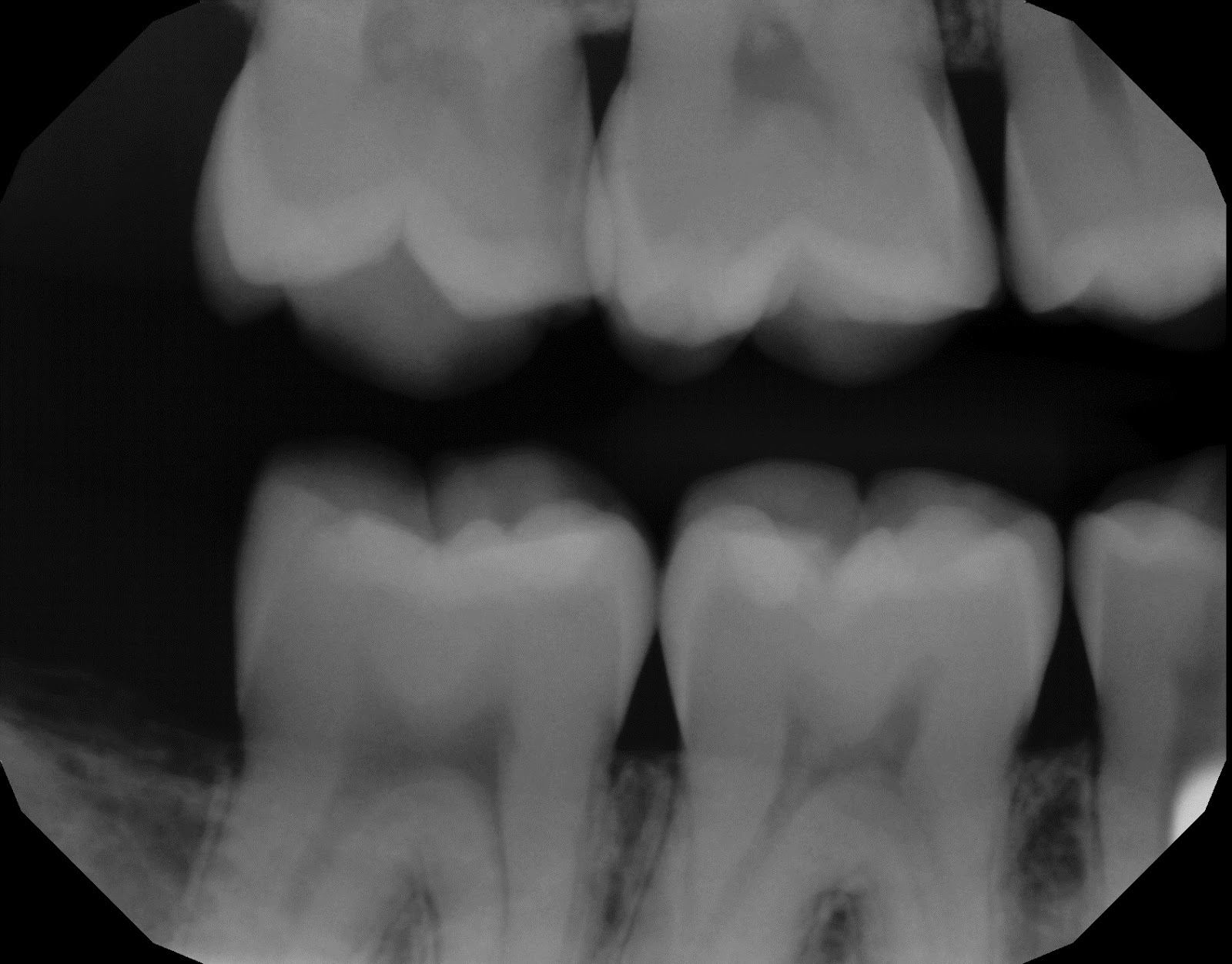


Dental x-rays are a case of "more than meets the eye."
Not only can the decay be bigger, it almost certainly will. Cavities on a tooth are always slightly larger in size than they would appear on an x-ray. For a standard dental radiograph, enamel has to loose about 40% of its mineral content before you can visualize the decay. In other words, the x-ray shows us a dark spot over the most demineralized areas, but the entire effected space will extend beyond this epicenter.
Furthermore, it is important to remember that an x-ray is only a 2D image. When looking at a radiograph, we can only make accurate judgements in one axis (from the part of the tooth closest to the throat to the part closest to the front of the mouth). We can gather some information on the dimensions from the cheek side to the tongue side, but it is less reliable. Thus, it can become difficult to judge the extent of decay as an entire 3D "space."