Viewing entries tagged
dental questions

Dental Questions: What do I do if I lose a temporary crown

Comment

Dental Questions: What do I do if I lose a temporary crown

Temporary crowns are not as developed or strong as permanent crowns because they are only intended to function for about two weeks. 

Temporary crowns are not as developed or strong as permanent crowns because they are only intended to function for about two weeks. 

Fortunately, losing a temporary crown is not a dental emergency. We primarily use them as space holders until the permanent restorations are ready. Even in the span of 2 weeks, the adjacent teeth can start to shift into the area created during the crown preparation. A temporary crown maintains space for the permanent crown, along with providing some chewing function and protection for the underlying tooth. If you lose a temporary crown, call our office and we will see you as soon as possible (typically the same afternoon/ following day). Remaking or recementing a temporary crown only takes about 20 minutes and usually does not require any anesthetic. Do not try to recement the crown on your own using drug store temporary dental cements. These products come with a number of risks, including permanently lodging the crown to the tooth or irritating the gums and periodontal space. Your teeth will not move significantly in the one to two days between losing your crown and having it replaced at our office. 

Comment

Dental Questions: Are water flossers a good substitute for dental floss?

Comment

Dental Questions: Are water flossers a good substitute for dental floss?

Water flossers (commonly known as “Waterpiks,” though that is a name brand) are typically marketed as being a convenient replacement for traditional dental floss. However, they can’t remove plaque and bacteria as thoroughly or completely as threaded floss. Overall, no product rivals the benefits of getting a physical object between the teeth and mechanically removing buildup. Still, a water flosser can be a great addition to your existing home care regimen. They are particularly good at removing packed-in food that your toothbrush can’t dislodge. Patients with gum disease will typically see the most benefit from a water flosser. The lowered height of gingival tissue allows the water to more effectively penetrate the periodontal space and remove debris.

Comment

Dental Questions: What is a “Same Day” Crown? How Does It Work?

Comment

Dental Questions: What is a “Same Day” Crown? How Does It Work?

Same day crowns are typically made by scanning the patient's tooth, designing a crown and milling it out of a ceramic/zirconia block.

Same day crowns are typically made by scanning the patient's tooth, designing a crown and milling it out of a ceramic/zirconia block.

    You may have seen advertisements for dental offices offering “same day” or “same visit” crowns. These services rely on a common engineering technology called CAD/CAM (computer automated design/ computer automated milling). During this procedure, your impression is taken digitally with a 3D scanner and uploaded to a special software that designs crowns. The dentist then finalizes the crown and sends it to a milling machine. After about an hour, the completed permanent restoration is ready to be finished and delivered to the patient. 

    On paper, this treatment sounds great and you may be wondering why we don’t offer it at our office. The reality is that these same-day crown systems have some serious drawbacks that we do not want to extend to our patients. For starters, an in-office milled crown is only as good as the time invested in it. Theoretically, we could scan your tooth, use a “generic” design, mill and deliver your crown in about 45 minutes. However, for these restorations to look and function properly, they need to be digitally adjusted, glazed and sintered in an oven. When done correctly, this process can take up to 2 hours; time you may not have to wait around! We much prefer utilizing the expertise of our local dental labs. Think of it this way: can a product produced in 45 minutes by a dental office really rival something that takes a master ceramist days to complete? 

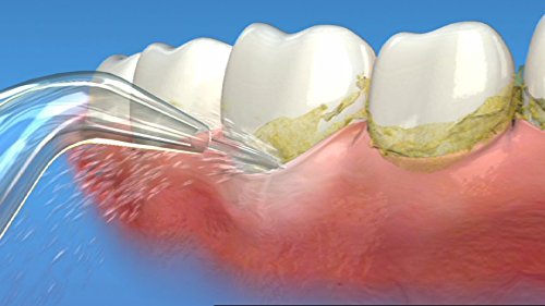
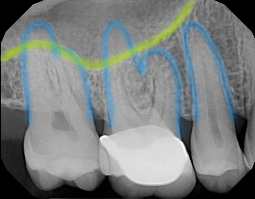
    Furthermore, the typical two week turnaround for a dental crown may seem like an inconvenience, but it actually serves a functional purpose. The unfortunate truth is that any time a dentist works on a tooth there is a (typically small) chance the nerve will become irritated and need a root canal. In the two weeks between preparing a tooth and cementing the crown, patients can usually tell if something is wrong with the nerve. Hence, we can provide a root canal before putting on the permanent crown. This can save a lot of headache in the future, particularly with the new, extremely hard zirconia crowns available. 

    Finally, using a CAD/CAM system severely limits the crown material choices available. There are no “one size fits all” dental materials (and anyone who tries to convince you otherwise is pulling your leg!). At our office, we treat each patient holistically and try to provide the best restorations for every tooth in its unique situation. There are a number of materials that these machines can’t use or can’t use well. With these milling units costing upwards of $100,000, many offices feel obligated or “locked-in” to providing CAD/CAM crowns, even when they are not the best option available. 

    As the technology currently stands, we are not comfortable offering in-office milled crowns to our patients. Our office feels that we get better results using a traditional dental lab to make our restorations. If you have any further questions or concerns on our crowns, how they are made or the materials we use, please give us a call!

Comment

Dental Questions: Why are my Gums Bleeding?

Comment

Dental Questions: Why are my Gums Bleeding?

thousandoaksfamilydentistry.com

Sometimes gums will bleed for obvious reasons, like cuts from flossing or abrasion from brushing. Other times, the root cause is less clear. The most common reason for gums to bleed randomly is due to calculus or tartar build up beneath the gum surface. Our office often sees patients that haven’t had their teeth professionally cleaned in a number of years. The number one complaint of these patients is that their gums bleed when brushing, when touched, or even while they are sleeping. A professional cleaning from a hygienist or dentist will remove the tartar, allow gums to heal and end the bleeding. 

Another common complaint is that gums bleed upon flossing. Interestingly, this is generally caused by not flossing enough. We recommend flossing or using some sort of hygiene instrument below the tooth contact once a day. This will help keep the gum tissue healthy and free of any plaque or tartar build up.

Comment

Dental Questions: Are sinus infections and pain on upper teeth related conditions?

Comment

Dental Questions: Are sinus infections and pain on upper teeth related conditions?

In this X-Ray, the floor of the maxillary sinus (yellow line) and the roots of the molars (blue lines) are highlighted. Note how close they are in proximity to each other. 

In this X-Ray, the floor of the maxillary sinus (yellow line) and the roots of the molars (blue lines) are highlighted. Note how close they are in proximity to each other. 

            The roots of the upper molars are frequently embedded in the floor of the maxillary sinus. As such, the nerves supplying the upper teeth can "pick up" pain signals from an inflamed or infected sinus. Since the size of the maxillary sinus grows with age, this phenomenon can become more prominent in later years. However, it is still important to consult a dentist about severe tooth pain or changes in the size or shape of your teeth and gums. This can be a potential sign of an infection that needs to be treated immediately. 

Comment

Dental Questions: Can decay on an x-ray look smaller than it actually is in the mouth?

Comment

Dental Questions: Can decay on an x-ray look smaller than it actually is in the mouth?

Dental x-rays are a case of "more than meets the eye."

Dental x-rays are a case of "more than meets the eye."


Not only can the decay be bigger, it almost certainly will. Cavities on a tooth are always slightly larger in size than they would appear on an x-ray. For a standard dental radiograph, enamel has to loose about 40% of its mineral content before you can visualize the decay. In other words, the x-ray shows us a dark spot over the most demineralized areas, but the entire effected space will extend beyond this epicenter.  

Furthermore, it is important to remember that an x-ray is only a 2D image. When looking at a radiograph, we can only make accurate judgements in one axis (from the part of the tooth closest to the throat to the part closest to the front of the mouth). We can gather some information on the dimensions from the cheek side to the tongue side, but it is less reliable. Thus, it can become difficult to judge the extent of decay as an entire 3D "space."

Comment

Dental Questions:  Can a dentist diagnose decay in the mouth that is not evident on an x-ray?

Comment

Dental Questions: Can a dentist diagnose decay in the mouth that is not evident on an x-ray?

Yes, it is absolutely possible to diagnose decay in the mouth from a visual exam with a mirror and explorer.  Decay on the biting surfaces of the teeth can often be hiding in a pit, groove or fissure of the outer enamel layer. On a visual exam, the area can look like a dark spot in the white of the enamel or an explorer can get stuck in the cavitation. The x-ray won’t show the decay in this area if is still primarily in the enamel layer. However, once decay spreads past this first layer of defense and lands in the internal dentin layer of the tooth, it will definitely show up on an xray.

Take a look at the following pictures, taken from the same patient in the same appointment:

In reviewing the radiographs, there is no apparent decay in this patient’s molars. There aren’t any suspicious shadows or defects in the enamel and the teeth generally look healthy. 

However, a visual examination reveals the beginnings of a cavity forming in the two maxillary molars. The geography of the tooth allows the decay to be hidden as the three dimensional object is translated into a 2D X-ray. Conversely, not all pit and fissure stains are cavities. Dentists can recognise unique shapes, patterns and texture changes in teeth that differentiate staining from active decay. This further highlights the importance of the entire dental exam to make a proper diagnosis. 

Comment