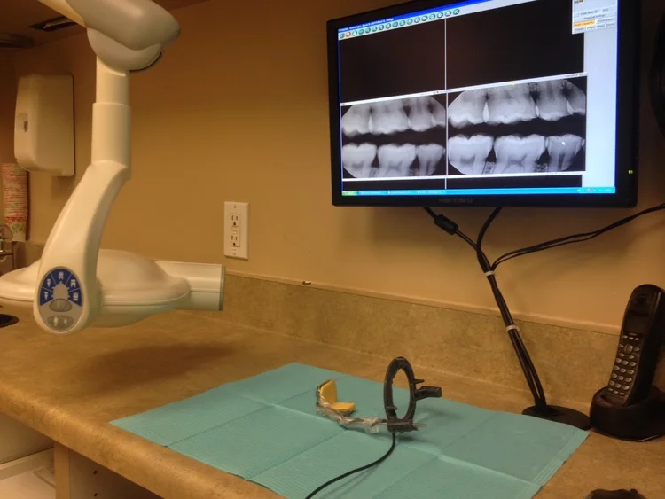
Most of our patients will remember the transition from film to digital dental x-rays with a positive stance. Gone are the days of sharp film holders, waiting for the developer and difficulties in duplication. But what are the biggest advantages of digital x-rays? Are they safe? How to they work? Read on to find out the true benefits behind this new technology!
Digital dental x-rays work on the same principles as modern digital cameras. In fact, our x-ray sensors use the same CMOS technology found in many modern DSLR, pocket and cell phone cameras. The main difference is that dental sensors are calibrated to react to x-ray radiation rather than the photons in visible light. Beyond that, the images are digitally decoded and rendered using much of the same technology as digital photography. The sensor itself contains no hazardous material and emits no radiation.
The two main principles that drive the digital revolution in dental x-rays are lower radiation and increased efficiency. "Exposing" a digital x-ray sensor requires less radiation than a similarly sized film, even when comparing the most sensitive film available. Digital images are also easier to manipulate, enlarge and change contrast, which allows us to gather more information off of a single picture. Finally, we can now judge the need for re-takes immediately, which translates to less waiting during your appointment.
Whenever discussing dental x-rays, the topic of safety always comes to light. We are here to assure our patients that dental radiography is extremely safe, with the benefits largely outweighing any drawbacks. Even after a full-mouth set of x-rays (taken about once every five years), you experience roughly the same amount of radiation as a trans-atlantic flight. Furthermore, four bitewings (taken about once every year to 18 months) require less radiation than spending a day outside in the sun. Particularly with our ultra sensitive digital sensors, your effective radiation exposure is negligible.
It is important to note that traditional film x-rays still play an important role in dentistry. Many practitioners still rely on film with excellent results and high patient satisfaction. This is particularly true of pediatric offices, where the thin profile and gentle flex of film allows dentists to get clearer images in smaller mouths. If you would like to know about digital x-rays or any other new trends in dental technology, please give our office a call!


